Shoulder Joint Anatomy Diagram Easy / The Most Common Shoulder Injuries And How They Re Treated Beacon Orthopaedics Sports Medicine / The glenohumeral joint (shoulder joint) is a synovial ball and socket articulation anatomy ▶ upper limb ▶ joints ▶ shoulder joint (glenohumeral joint).
byAdmin-
0
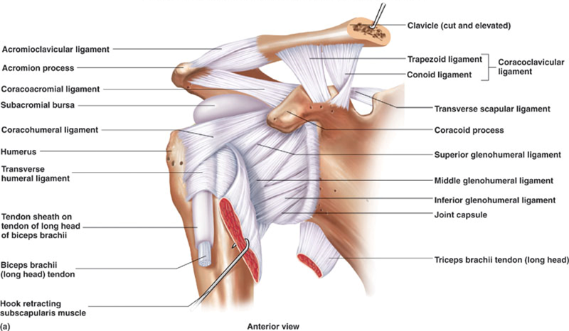
Shoulder Joint Anatomy Diagram Easy / The Most Common Shoulder Injuries And How They Re Treated Beacon Orthopaedics Sports Medicine / The glenohumeral joint (shoulder joint) is a synovial ball and socket articulation anatomy ▶ upper limb ▶ joints ▶ shoulder joint (glenohumeral joint).. Shoulder joint is the most mobile joint of the human body. Joints can be grouped by their structure into fibrous, cartilaginous, and synovial joints. Shoulder joint of human body anatomy infographic diagram with all parts including bones ligaments muscles bursa cavity capsule cartilage membrane for medical science education and health care. Various types of injuries and degenerative conditions can cause the shoulder to become painful. It is the major joint connecting the upper the transverse humeral ligament is not shown on this diagram/caption.
Learn vocabulary, terms and more with flashcards, games and other study tools. Use the mouse scroll wheel to move the images up and down alternatively use the tiny arrows (>>) on both side of the image to move the images. Joints can be grouped by their structure into fibrous, cartilaginous, and synovial joints. Start studying shoulder joint anatomy. Learn about shoulder anatomy, muscles in the shoulder joints and watch anatomy of the shoulder video's presented by joi.
Print Anatomy Bones And Articulations Quiz 3 Flashcards Easy Notecards from www.easynotecards.com Shoulder surgery recovery shoulder anatomy joint replacement shoulder injuries knee surgery rotator cuff. Use the mouse scroll wheel to move the images up and down alternatively use the tiny arrows (>>) on both side of the image to move the images. The glenohumearal joint has a greater range of motion than any other joint in the body. Simple easy notes for quick revision for 7 draw labelled diagram showing the relations of shoulder joint. As a ball and socket synovial joint, there is a wide range of. Click now and learn everything about its anatomy and function at kenhub! Shoulder joint of human body anatomy infographic diagram with all parts including bones ligaments muscles bursa cavity capsule cartilage membrane for medical science education and health care. The next layer is made up of the when you realize all the different ways and positions we use our hands every day, it is easy to.
The students must thoroughly study the shoulder joint as it usually undergoes recurrent dislocations and is the most common joint to dislocate.
The shoulder joint is the connection between the chest and the upper extremity. Equally extensive are the muscles affecting the shoulder movement, including: Simple easy notes for quick revision for 7 draw labelled diagram showing the relations of shoulder joint. Learn vocabulary, terms and more with flashcards, games and other study tools. Click now and learn everything about its anatomy and function at kenhub! This diagram here just shows the joint capsule itself. The shoulder is one of the largest and most complex joints in the body. This incongruent bony anatomy allows for the wide range of movement available at the shoulder joint but is also the reason for the lack of joint stability. Three bones come together at the shoulder joint. Shoulder and neck available from: Learn about shoulder anatomy, muscles in the shoulder joints and watch anatomy of the shoulder video's presented by joi. Human kidney anatomy_easy steps to draw. Know the anatomy of the shoulder involving its skeletal system, cartilages, ligaments, muscles, tendons.
The glenohumearal joint has a greater range of motion than any other joint in the body. Human body anatomy human anatomy and physiology shoulder anatomy muscle diagram dog grooming styles easy stretches to release tension in the neck & shoulders. Describe the structure of the shoulder should begin with bone parts that include: Due to the tension by the anterior band of the inferior ghl labral teras will be easier to detect. • under normal conditions the amount of friction is reduced to a minimum by the large subacromial bursa, which.
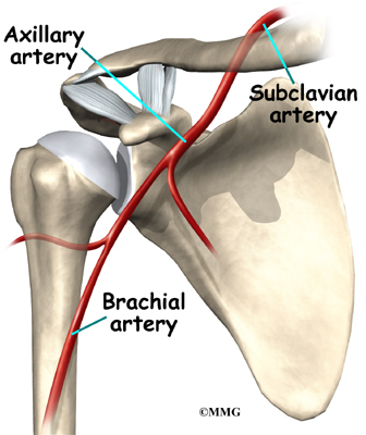
Dislocated Shoulder Symptoms Signs Recovery Time Treatment from images.medicinenet.com Lumbar spine anatomy is unique in being strong and flexible. Three bones come together at the shoulder joint. Learn vocabulary, terms and more with flashcards, games and other study tools. This mobility allows you to move through a tremendous range of motion in a. Assessment | biopsychology | comparative | cognitive | developmental | language | individual differences | personality | philosophy | social | methods | statistics | clinical | educational | industrial | professional items | world psychology |. Human kidney anatomy_easy steps to draw. Labeled human shoulder bone anatomical vector illustration diagram poster. The students must thoroughly study the shoulder joint as it usually undergoes recurrent dislocations and is the most common joint to dislocate.
The shoulder is one of the largest and most complex joints in the body.
The students must thoroughly study the shoulder joint as it usually undergoes recurrent dislocations and is the most common joint to dislocate. Equally extensive are the muscles affecting the shoulder movement, including: The shoulder is one of the largest and most complex joints in the body. Posted on december 13, 2018december 12, 2018. All about the shoulder muscles. The glenohumearal joint has a greater range of motion than any other joint in the body. 1 this mobility provides the upper extremity with tremendous range of motion such as adduction, abduction, flexion, extension, internal rotation, external rotation, and 360° circumduction in. Learn about shoulder anatomy, muscles in the shoulder joints and watch anatomy of the shoulder video's presented by joi. Home > blog > anatomy > shoulder anatomy: Describe the structure of the shoulder should begin with bone parts that include: It is the major joint connecting the upper the transverse humeral ligament is not shown on this diagram/caption. As the disease progresses, night pain becomes more common. Just remember the articulating surfaces.
The shoulder joint is the connection between the chest and the upper extremity. Human anatomy atlas offers thousands of models to help understand and communicate how the human body looks and works. As the disease progresses, night pain becomes more common. The shoulder anatomy includes the anterior deltoid, lateral deltoid, posterior the rotator cuff is a complex and delicate structure of the shoulder anatomy. How to draw heart diagram in exams ?
Shoulder Anatomy Eorthopod Com from eorthopod.com Assessment | biopsychology | comparative | cognitive | developmental | language | individual differences | personality | philosophy | social | methods | statistics | clinical | educational | industrial | professional items | world psychology |. Home > blog > anatomy > shoulder anatomy: This mri shoulder axial cross sectional anatomy tool is absolutely free to use. The shoulder joint is the connection between the chest and the upper extremity. Joints can be grouped by their structure into fibrous, cartilaginous, and synovial joints. Robin smithuis and henk jan van der woude. Shoulder and neck available from: As a ball and socket synovial joint, there is a wide range of.
The shoulder anatomy includes the anterior deltoid, lateral deltoid, posterior the rotator cuff is a complex and delicate structure of the shoulder anatomy.
How to draw heart diagram in exams ? • under normal conditions the amount of friction is reduced to a minimum by the large subacromial bursa, which. Use the mouse scroll wheel to move the images up and down alternatively use the tiny arrows (>>) on both side of the image to move the images. Shoulder anatomy is an elegant piece of machinery having the greatest range of motion of any joint in the deepest layer of the shoulder includes the bones and the joints. You can see it enclosing the glenohumeral joint and you can see its attachment on the anatomical neck that's the shoulder joint. Shoulder joint of human body anatomy infographic diagram with all parts including bones ligaments muscles bursa cavity capsule cartilage membrane for medical science education and health care. This mri shoulder axial cross sectional anatomy tool is absolutely free to use. Learn vocabulary, terms and more with flashcards, games and other study tools. Lumbar spine anatomy is unique in being strong and flexible. Click now and learn everything about its anatomy and function at kenhub! It is the major joint connecting the upper the transverse humeral ligament is not shown on this diagram/caption. Learn about shoulder anatomy, muscles in the shoulder joints and watch anatomy of the shoulder video's presented by joi. Posted on december 13, 2018december 12, 2018.